Colorante catiónico que se acumula selectivamente en las mitocondrias, probablemente a través del gradiente de potencial de la membrana mitocondrial.
Descripción
MitoLite™ Red es un tinte catiónico que se acumula selectivamente en las mitocondrias, probablemente a través del gradiente de potencial de la membrana mitocondrial. El indicador mitocondrial es un compuesto hidrofóbico que penetra fácilmente en las células vivas intactas y queda atrapado en las mitocondrias después de que ingresa a las células. Este indicador mitocondrial fluorescente se retiene en las mitocondrias durante mucho tiempo ya que el indicador lleva un grupo de retención celular. Esta característica clave aumenta significativamente su eficacia de tinción.
El protocolo de etiquetado es sólido y requiere un tiempo mínimo de manipulación. Puede adaptarse fácilmente a una amplia variedad de plataformas de fluorescencia, como ensayos en microplaca, inmunocitoquímica y citometría de flujo. Es adecuado para células proliferantes y no proliferativas, y se puede utilizar tanto para células en suspensión como adherentes.
| Catalogo | Producto | Presentación |
|---|---|---|
| AAT-22677 | MitoLite™ Red FX600 | 500 pruebas |
Importante: Solo para uso en investigación (RUO). Almacenamiento: Congelación (< -15 °C). Minimizar la exposición a la luz.
Espectro
Abrir en Advanced Spectrum Viewer

Propiedades espectrales
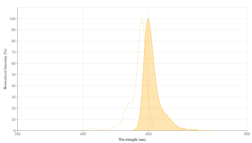
| Exitación | 580 |
| Emisión | 598 |
Imagen

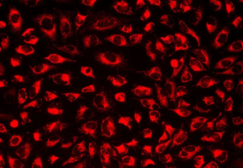
Figura 1. Imagen de células U2OS teñidas con MitoLite™ Red FX600 en una placa de 96 pocillos de pared negra/fondo transparente.

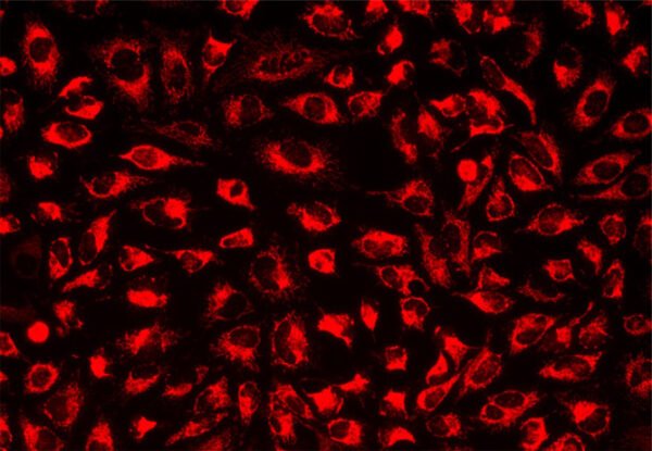
Figura 2. Retención celular de MitoLite™ Red FX600 en células HeLa. Las células HeLa vivas se tiñeron con MitoLite™ Red FX600 en buffer HH durante 30 min a 37oC. Las células se lavaron 3 veces con buffer HH y luego se cultivaron continuamente en medio fresco durante 2 días. Las imágenes inmediatamente después de la tinción (Día 0), ~24 (Día 1) y 48 horas (Día 2) después de la tinción se tomaron usando un microscopio de fluorescencia con un juego de filtros Texas Red®.
Productos Relacionados
| MitoLite™ Red CMXRos |
| MitoLite™ Blue FX490 |
| MitoLite™ Green EX488 |
| MitoLite™ Orange FX570 |
| MitoLite™ Deep Red FX660 |
| MitoLite™ Orange EX405 |
| MitoLite™ NIR FX690 |
| MitoLite™ Green FM |
Bibliografía
Chitosan Oligosaccharides Alleviate H2O2-stimulated Granulosa Cell Damage via HIF-1$\alpha$ Signaling Pathway
Authors: Yang, Ziwei and Hong, Wenli and Zheng, K and Feng, Jingyuan and Hu, Chuan and Tan, Jun and Zhong, Zhisheng and Zheng, Yuehui
Journal: Oxidative Medicine and Cellular Longevity (2022)
Co-delivery of VP-16 and Bcl-2-targeted antisense on PEG-grafted oMWCNTs for synergistic in vitro anti-cancer effects in non-small and small cell lung cancer
Authors: Heger, Zbynek and Polanska, Hana and Krizkova, Sona and Balvan, Jan and Raudenska, Martina and Dostalova, Simona and Moulick, Amitava and Masarik, Michal and Adam, Vojtech
Journal: Colloids and Surfaces B: Biointerfaces (2017): 131–140
Inhibition of heme oxygenase-1 enhances the chemosensitivity of laryngeal squamous cell cancer Hep-2 cells to cisplatin
Authors: Lv, Xin and Song, Dong-mei and Niu, Ying-hao and Wang, Bao-shan
Journal: Apoptosis (2016): 489–501
Effective two-photon excited photodynamic therapy of xenograft tumors sensitized by water-soluble bis (arylidene) cycloalkanone photosensitizers
Authors: Zou, Qianli and Zhao, Hongyou and Zhao, Yuxia and Fang, Yanyan and Chen, Defu and Ren, Jie and Wang, Xiaopu and Wang, Ying and Gu, Ying and Wu, Feipeng
Journal: Journal of medicinal chemistry (2015): 7949–7958
Melatonin promotes adipogenesis and mitochondrial biogenesis in 3T3-L1 preadipocytes
Authors: Kato, Hisashi and Tanaka, Goki and Masuda, Shinya and Ogasawara, Junetsu and Sakurai, Takuya and Kizaki, Takako and Ohno, Hideki and Izawa, Tetsuya
Journal: Journal of Pineal Research (2015): 267–275
Referencias
Ver todas las 70 referencias: Citation Explorer
Quantification of carbonylated proteins in rat skeletal muscle mitochondria using capillary sieving electrophoresis with laser-induced fluorescence detection
Authors: Feng J, Arriaga EA.
Journal: Electrophoresis (2008): 475
Calcium, mitochondria and apoptosis studied by fluorescence measurements
Authors: Roy SS, Hajnoczky G.
Journal: Methods (2008): 213
Fluorescence imaging of mitochondria in yeast
Authors: Swayne TC, Gay AC, Pon LA.
Journal: Methods Mol Biol (2007): 433
A fluorescence assay for peptide translocation into mitochondria
Authors: Martinez-Caballero S, Peixoto PM, Kinnally KW, Campo ML.
Journal: Anal Biochem (2007): 76
Fast electrophoretic analysis of individual mitochondria using microchip capillary electrophoresis with laser induced fluorescence detection
Authors: Duffy CF, MacCraith B, Diamond D, O’Kennedy R, Arriaga EA.
Journal: Lab Chip (2006): 1007
Discrimination of depolarized from polarized mitochondria by confocal fluorescence resonance energy transfer
Authors: Elmore SP, Nishimura Y, Qian T, Herman B, Lemasters JJ.
Journal: Arch Biochem Biophys (2004): 145
A fluorescence-based technique for screening compounds that protect against damage to brain mitochondria
Authors: Kristian T, Fiskum G.
Journal: Brain Res Brain Res Protoc (2004): 176
Determination of the cardiolipin content of individual mitochondria by capillary electrophoresis with laser-induced fluorescence detection
Authors: Fuller KM, Duffy CF, Arriaga EA.
Journal: Electrophoresis (2002): 1571
Fluorescence imaging of metabolic responses in single mitochondria
Authors: Nakayama S, Sakuyama T, Mitaku S, Ohta Y.
Journal: Biochem Biophys Res Commun (2002): 23
Visualisation of mitochondria in living neurons with single- and two-photon fluorescence laser microscopy
Authors: Dedov VN, Cox GC, Roufogalis BD.
Journal: Micron (2001): 653
Application notes (en Ingles)
A Novel Fluorescent Probe for Imaging and Detecting Hydroxyl Radical in Living Cells
Abbreviation of Common Chemical Compounds Related to Peptides
Annexin V
Bright Tide Fluor™-Based Fluorescent Peptides and Their Applications In Drug Discovery and Disease Diagnosis
Calcein


